Patello Femoral Instability
Performed by – Khalid Al-Hourani, Alastair Davidson, Reza Mansouri, Zuhair Nawaz, Andrew Perry, James Singleton
Instability of the patella (kneecap) can present with pain or in more extreme circumstances dislocation. There are many reasons why the patella may not work well and some will require surgery to correct them.
In the first instance a careful clinical examination is required together with plain X rays and possibly an MRI scan. In some cases, a CT scan may also be required.
The majority of cases will be treated with physiotherapy in the first instance to strengthen the muscles of the thigh and improve the tracking of the patella. This will improve the symptoms in the majority of patients and apart from maintaining the muscles, no further treatment is required.
In some patients there are anatomical abnormalities that mean physiotherapy is unlikely to resolve the problem. Sometimes the patella can be too high, the groove on the front of the femur (thigh bone) which the patella passes down may be too shallow, the attachment of the tendon to the front of the tibia (tibial tubercule) can be too lateral and the ligaments on the inside of the patella can be weak or damaged after a dislocation. All of these make dislocation more likely to occur in the future.
Surgical treatment will be directed at the problem or problems as more than one is frequently present.
If the patella is too high or the tibial tubercule too laterally placed, the tubercule can be moved inwards or downwards to correct the problem. This is called a tibial tubercule osteotomy.
If the ligaments on the inside of the patella are weak or damaged they can be reconstructed using a tendon graft to make them strong again. This is called a medial patella femoral ligament reconstruction.
If the groove on the front of the femur down which the patella passes is too shallow it can be deepened with a procedure called a trochleoplasty.
Patello Femoral surgery
Generally, you will be in hospital overnight. The surgery is done under a general anaesthetic and local anaesthetic is injected around the wound for pain relief after the operation.
Surgery can generally be done using several small transverse incisions, about 2cm long, which will heal better than a single vertical incision.
You will need to be in a leg immobiliser splint for a period after surgery to support the knee but in most cases you will be able to take full weight on the leg early on. Physiotherapy is very important and in most cases the leg immobiliser splint can be removed when you regain a straight leg raise, generally in 2-3 weeks. By 6 weeks you should be able to walk normally. The exception to this is surgery to move the tibial tubercule downwards when you will need a leg immobiliser splint and crutches for 6 weeks while the bone heals.
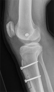
- X Ray after surgery to reconstruct the medial patella femoral ligaments and move the tibial tubercle distally
- AP X Ray after surgery to reconstruct the medial patello femoral ligaments and move the tibial tubercle distally
- X Ray after surgery to reconstruct the medial patella femoral ligaments and move the tibial tubercle distally
- Lateral X Ray after surgery to reconstruct the medial patello femoral ligaments and move the tibial tubercle distally